Radius Bone Labelled Diagram / humerus_labeled - We have some clues that ulna is thinner and longer than radius.. Study guide for students and teachers. The radius is considered the most commonly fractured bone in the human body, with distal radius fractures being the most common form of radial. The radius and ulna together constitute the forearm. Human anatomy diagrams show internal organs bone diagram barca fontanacountryinn com. 12 photos of the labelled diagram of radius bone.
The biceps originate near the shoulder joint and insert into the radial tuberosity on the upper part of the radius, near the elbow joint. Its upper concave surface articulates with the. Radius and ulna bones anatomy introduction. Study guide for students and teachers. The radius is considered the most commonly fractured bone in the human body, with distal radius fractures being the most common form of radial.
Detailed Human Skeleton Diagrams - Health, Medicine and ... from i.pinimg.com Roughened surface where the biceps brachii inserts. Radius bone is a photograph by asklepios medical atlas which was uploaded on august 3rd, 2016. Skeletal system diagrams are illustrations of the human skeleton. Bone structure diagram wiring diagrams click. Proximal radius (head, neck and tuberosity). Thats the way i remembered which bone is located in anatomy class. The radius bone is the lateral bone of the forearm, and is homologous with the tibia of the lower limb. Even my bio teacher get confused.
Radius, in anatomy, the outer of the two bones of the forearm when viewed with the palm facing forward.
Roughened surface where the biceps brachii inserts. The radius is the bone which is. Learn radius and ulna anatomy with these fun quizzes and diagrams. It extends from the lateral side of the elbow to the thumb side of the wrist and runs parallel to the ulna. The biceps originate near the shoulder joint and insert into the radial tuberosity on the upper part of the radius, near the elbow joint. There are different features on each bone that also can help distinguish between the. Radius along with ulna connects elbow to forearm. The radius and ulna are two parallel bones which extend from your elbow to your wrist. The radius is a long bone in the forearm. Short video describing the skeletal structures of the radiusstructures identified:headneckradial tuberositystyloid process of the radiusulnar notch. Radius, in anatomy, the outer of the two bones of the forearm when viewed with the palm facing forward. In the medial surface, there. It lies laterally and parallel to ulna, the second of the forearm bones.
Short video describing the skeletal structures of the radiusstructures identified:headneckradial tuberositystyloid process of the radiusulnar notch. Cheek bone (zygoma) upper jaw (maxilla). The radius bone is a long horizontal bone present in the forearm and is also called the radial bone. We have some clues that ulna is thinner and longer than radius. Radial tuberosity (tuberositas radii) is an oval elevation on the proximal, medioanterior margin of the radius.
Pin by Lori Stewart on College | Anatomy bones, Human ... from i.pinimg.com The radius is the bone which is. The radius bone is a long horizontal bone present in the forearm and is also called the radial bone. In humans it is shorter than the other bone of the forearm, the ulna. Radius and ulna bones anatomy introduction. It extends from the lateral side of the elbow to the thumb side of the wrist and runs parallel to the ulna. Roughened surface where the biceps brachii inserts. The lateral side projects distally as the styloid process. There are different features on each bone that also can help distinguish between the.
It is simulated by using a 12 kg/cm servo motor with gears.
Radius ulna bones ulnar notch anterior anatomy markings head forearm wrist hand limb upper pronation pivot supination during around. 12 photos of the labelled diagram of radius bone. The radial head of the flexor digitorum superficialis takes origin from the anterior oblique line and upper part of the anterior border. Bone structure diagram wiring diagrams click. 720 x 904 jpeg 49 кб. Lower jaw (mandible) collar bone. The two bones play only secondary roles at their opposing joints. It is located laterally and extends from the elbow to the wrist. Learn everything about the anatomy of radius and ulna with our articles, video tutorials, labeled diagrams, and quizzes. The ulna is usually slightly longer than the radius, but the radius is thicker. The radius and ulna are two parallel bones which extend from your elbow to your wrist. Radius bone is a photograph by asklepios medical atlas which was uploaded on august 3rd, 2016. The lateral side projects distally as the styloid process.
Learn radius and ulna anatomy with these fun quizzes and diagrams. The photograph may be purchased as wall art, home decor, apparel, phone cases, greeting cards, and more. The bones mentioned in each human skeleton chart are: The radius and ulna together constitute the forearm. In the medial surface, there.
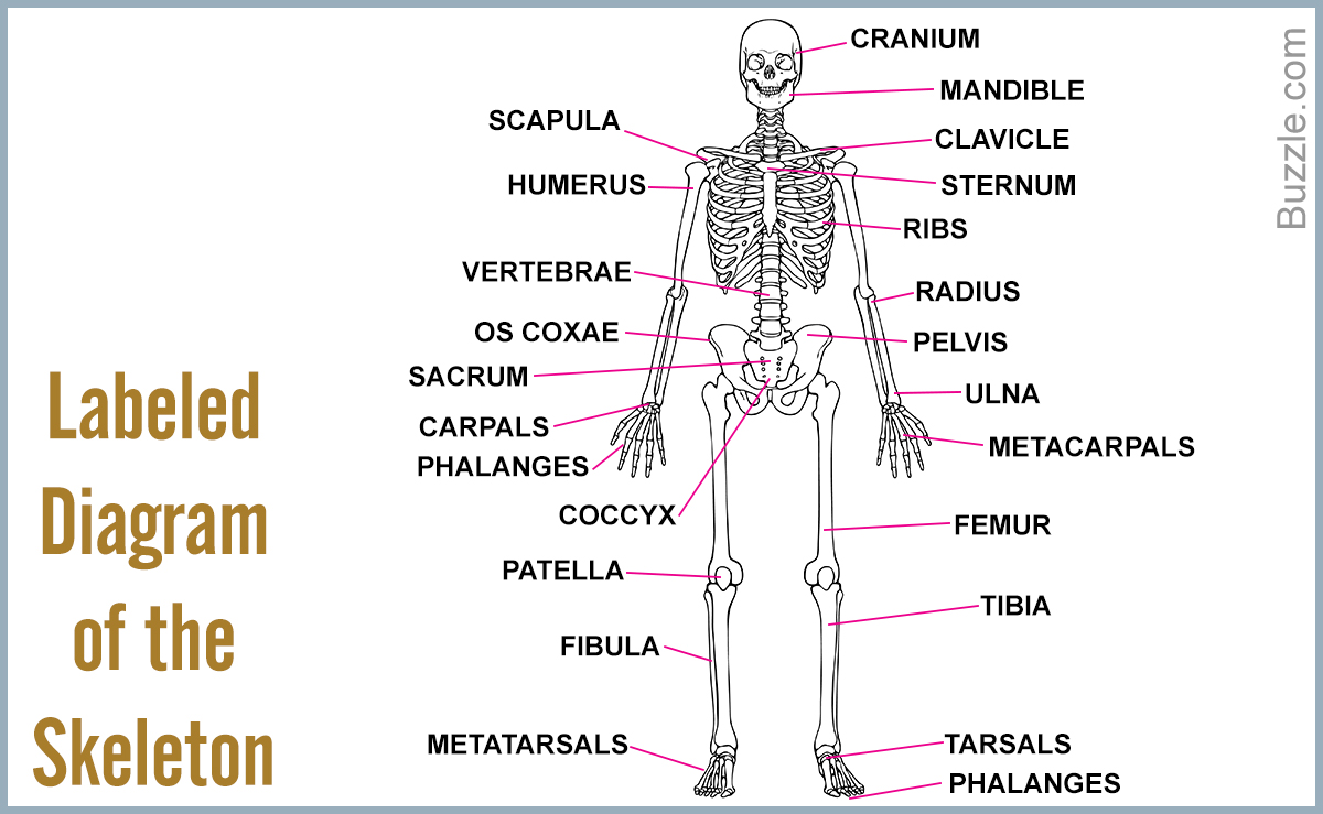
A List of Bones in the Human Body With Labeled Diagrams ... from media.buzzle.com The bones shown in the chest and hip region in the labeled human skeleton diagram are the ribs humerus is located in the upper arm. Short video describing the skeletal structures of the radiusstructures identified:headneckradial tuberositystyloid process of the radiusulnar notch. The radial head of the flexor digitorum superficialis takes origin from the anterior oblique line and upper part of the anterior border. There are different features on each bone that also can help distinguish between the. The radius bone is the lateral bone of the forearm and is homologous with the tibia of the lower limb. In humans it is shorter than the other bone of the forearm, the ulna. It is simulated by using a 12 kg/cm servo motor with gears. The radius is the home for a few muscles' insertion points.
The radius bone is a long horizontal bone present in the forearm and is also called the radial bone.
The elbow articulates in 4 places radial tuberosity: All land vertebrates have this bone. Human anatomy diagrams show internal organs bone diagram barca fontanacountryinn com. This ulnar view labelled illustration is from 'asklepios atlas of the human anatomy'. Learn everything about the anatomy of radius and ulna with our articles, video tutorials, labeled diagrams, and quizzes. Radial tuberosity (tuberositas radii) is an oval elevation on the proximal, medioanterior margin of the radius. In its distal part, the radial shaft expands to form a rectangular end. Proximal radius (head, neck and tuberosity). Lower jaw (mandible) collar bone. We have some clues that ulna is thinner and longer than radius. The radius and ulna are the two bones of the forearm. Bone structure diagram wiring diagrams click. Its upper concave surface articulates with the.
Each bone is a complex living organ that is made up of many cells, protein fibers, and minerals labelled radius bone. Labeled ulna and radius » diagram text anterior labeled ulna and radius anatomy of the ulna and radius bones print options de labeled ulna.


0 Komentar